Nancy Kanwisher and Rosa Lafer-Sousa in The Neuroanatomy Lesson
Ever wonder which areas of your brain “turn on” when you see faces or think about other people’s thoughts? Using a scanning method called fMRI, researchers like MIT neuroscientist Nancy Kanwisher can pinpoint where brain cells carry out these and other mental functions.
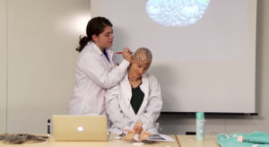
Such digital images are powerful, but Kanwisher, in a recent installment of her video series “Nancy’s Brain Talks,” offers more. “It’s kind of hard to tell [where these particular brain regions are] with all this damn hair in the way,” she says, and proceeds to shave herself bald. A “scalp neuro-artist” (graduate student Rosa Lafer-Sousa) then draws a pizza-sized cortex on her head, coloring in the regions that process language, bodies, and places.
With the help of fMRI, researchers like Kanwisher will soon learn whether there are special brain circuits for hearing the sounds of speech or appreciating a melody. “It’s a super exciting time to be investigating the human mind and brain,” she says in the video. “Even if you don’t shave your head.”
Explore Professor Kanwisher’s research in the Open Access Articles collection in DSpace@MIT, where it is openly accessible to the world.
Since the MIT faculty established their Open Access Policy in March 2009 they have made thousands of research papers freely available to the world via DSpace@MIT. To highlight that research, we’re offering a series of blog posts that link news stories about scholars’ work to their open access papers in DSpace.